
WHATS NEW IN OI
 https://pmc.ncbi.nlm.nih.gov/articles/PMC13092026/pdf/main.pdf
march 2026
Current Concept Review
What's New in Osteogenesis Imperfecta
Maegen Wallace, MD, MBA, FAAOS, FAOA1,*; Bryan Menapace, MD, MBA2;
Christina Herrero, MD3; Jeanne M. Franzone, MD, FAAOS, FAOA4
1 Phoenix Children's Hospital, Phoenix, AZ, USA
2 Shriners Children's Portland, Portland, OR, USA
3 RWJBarnabas Health, New Brunswick, NJ, USA
4 Nemours Children's Hospital Delaware, Wilmington, DE, USA
A R T I C L E I N F O
Keywords:
Osteogenesis imperfecta
Bisphosphonates
Telescoping rods
Scoliosis
A B S T R A C T
Osteogenesis imperfecta (OI) is a diverse group of genetic disorders mainly caused by pathogenic variants in COL1A1 and COL1A2 that interfere with type I collagen production, resulting in bone fragility and multisystem issues. Since 2019, the understanding and treatment of OI have improved considerably. Advances in molecular genetics have broadened the OI classification to 22 types, with new variants affecting pathways beyond collagen synthesis, including osteoblast differentiation and bone mineralization. Bisphosphonates continue to be the primary pharmacologic treatment, effectively reducing fracture risk, and increasing bone mineral density, although recent studies highlight delayed osteotomy healing and some differences among specific agents. Newer therapies—including denosumab, antisclerostin antibodies such as setrusumab, and mesenchymal stem cell
transplantation—show promise but need further validation through randomized trials. Overall, nonoperative care, including good nutrition, vitamin D supplementation, personalized physical therapy, and orthotic support, remains essential for improving function and preventing fractures. Intramedullary stabilization with telescoping rods has become the standard for both fracture fixation and deformity correction, providing better long-term outcomes and lower reoperation rates compared to static rods. Advances in surgical planning highlight the importance of precise rod alignment, avoiding cortical stress shielding, and protecting the periosteum to promote healing. Spinal deformities, including scoliosis and basilar invagination, are increasingly managed through multidisciplinary monitoring, bisphosphonate therapy, and refined surgical strategies that prioritize stability over maximum correction. Optimal outcomes in OI require coordinated multidisciplinary care involving or-
thopaedic surgeons, medical colleagues, physical therapists, and other specialists. Ongoing research continues to refine medical and surgical methods to improve function, quality of life, and long-term skeletal health for children with OI.
Key Concepts:
(1) Multidisciplinary care is vital in caring for children with OI.
(2) Medical optimization through nutrition, medications like bisphosphonates, vitamin D supplementation, and physical therapy is important for both preoperative and postoperative children with OI.
(3) Intramedullary stabilization with telescoping or nontelescoping nails to protect the entire length of the bone is the mainstay of surgical treatment.
Introduction
The field of rare bone diseases, including osteogenesis imperfecta (OI), has advanced due to new research in recent years. This review aims to update readers on current developments in the medical and surgical management of OI, rather than providing a comprehensive historical overview of the condition. Literature was identified through PubMed searches, as well as abstracts from the Pediatric Orthopaedic Society of North America (POSNA) meetings, the International Conference for
Children's Bone Health (ICCBH) 2024, and the International Conference on OI (ICOI) 2025.
The OI is a heterogeneous group of genetic disorders characterized by bone fragility and abnormal connective tissue, commonly called “brittle bone disease.” The most common genetic variants occur in
COL1A1 and COL1A2, although many other variants affecting collagen synthesis, osteoblast development, or extracellular matrix pathways have been identified. Clinical features vary widely from perinatal death to milder forms mainly marked by increased fracture risk. The original Sillence classification described four clinical types before the understanding of the genetic cause [1]. The genetic cause of OI was identified in the early 1980s [2,3]. Type V OI was later defined by
Glorieux et al. in 2000 [4], and in 2006, the first recessive form of OI was identified [5]. As new gene variants are discovered, the classification system has grown to include more than 20 types. Table 1 summarizes these forms of OI.
The OI is a multisystem condition with many extra-skeletal mani-festations in the bone, muscle, tendon, sclerae, blood vessels, and the gastrointestinal tract [6]. Common signs include blue sclera, hearing loss, dental and craniofacial differences, skeletal muscle weakness, and cardiovascular issues [7,8]. Effective management, therefore, requires a multidisciplinary approach involving [9–13] medical bone health providers (orthogenetics, endocrinology, or nephrology), orthopaedic surgery, physical and occupational therapy, pain management, psy- chology, audiology, cardiology, social work, and nutritionists. Opti-mizing medical care is crucial to improving orthopaedic outcomes in OI.
Treatment options include nutritional support, medications affecting bone metabolism, and ongoing monitoring of the skeletal, pulmonary, auditory, and cardiovascular systems [14]. Adequate calcium and vitamin D intake are vital, and careful attention to diet is recom- mended to prevent poor growth in infancy and excessive weight in older children. Bisphosphonates remain the primary pharmacologic
treatment as they decrease fracture risk, increase bone mineral density (BMD), alleviate chronic bone pain, and assist in prophylactic rodding of long bones [15,16].
Bisphosphonates
Pamidronate and Zoledronate are the most common bisphospho-nates used in children with OI and may be started in infancy includingin the neonatal intensive care unit (NICU) for infants with moderate
to severe disease. These medications inhibit osteoclast-mediated boneresorption, allowing increased bone mass and strength during growth.The most common adverse reaction is a transient febrile response after the first infusion, though the medications are generally well tolerated otherwise. Bone health is usually monitored with serial dual-energy x-ray absorptiometry (DEXA) scans during childhood to observe changes in bone density. Bisphosphonate therapy often continues until skeletal maturity or until bone density normalizes.
Most clinicians do not delay treatment after fractures, although some recommend delaying therapy after planned osteotomies or spinal fusion until early radiographic signs of healing appear. Concerns
have been raised about atypical fractures in patients on long-term therapy, but current evidence suggests these fractures are more closely linked to disease severity than to bisphosphonate exposure
A systematic review published in 2025 evaluated fracture rates, lumbar spine bone mineral density (BMD), and safety outcomes asso-ciated with bisphosphate therapy in OI. Overall treatment significantly improved BMD and reduced fractures, though outcomes varied among individual agents. The authors suggested that neridronate and olpadr-onate may offer favorable efficacy and tolerability profiles, while pamidronate and alendronate require careful monitoring for adverse effects. Oral agents were associated with gastrointestinal symptoms, whereas IV treatments were more commonly associated with flu-like reactions [19].
A 2024 retrospective study examined the impact of bisphosphonate therapy on osteotomy healing in children with moderate to severe OI. Among nine osteotomies performed in six patients, those receiving bisphosphonates experienced significantly long healing times (10.4 months) compared with controls (3.5 months). Radiographs demon-strated limited callus formation and periosteal new bone, suggesting a healing pattern resembling primary bone healing. These findings raise concerns regarding mechanical strength and delayed weight-bearing in this population [20].
Other medications
Denosumab, a monoclonal antibody targeting RANK ligand, has been used off-label in children with OI, although evidence remains limited. The most concerning adverse effect is rebound hypercalcemia after discontinuation, with some reports of hypercalcemic crisis occurring in up to 14.3% of patients
Another emerging therapy involves antisclerostin antibodies such as setrusumab. Phase II trial data presented at ICCBH in 2024 included twenty-four children with OI (71% type I and 29% type III). The
medication was found to be safe and showed a mean increase in spine BMD from baseline to month 3 of 9.1% and to month 6 of 12.8% (P > .05 vs baseline) [23]. A phase 3 data lock and analysis are currently un-derway, and at the time of this manuscript, there are no published data to report.
Mesenchymal stem cell (MSC) transplantation is another investiga-tional approach. A 2025 systematic review identified nine studies involving twelve patients treated with bone marrow–derived MSC s
(BMSCs) or human fetal MSCs (hfMSCs). Reported outcomes included improved bone mineralization, reduced fracture rates, and enhanced growth. Engraftment rates ranged from 1 to 7%, and adverse effects were generally temporary. Larger randomized trials are needed before widespread clinical adoption
Nonoperative treatment
Nonoperative treatment is crucial for preventing fractures. Early intervention therapy can help children with OI who are delayed in reaching motor milestones. It is essential for therapists to understand the
pathology of OI so they can customize therapies and interventions based on each child's disease severity and safety needs. Often, multidisci- plinary assessments including physical therapy (PT), occupational therapy (OT), aquatic therapy, and behavioral and emotional support are most effective for developing long-term plans. It is important for both family members and therapists to recognize the need to balance bone protection with muscle development. Muscle weakness can lead to
further reduced bone strength, making these challenges worse.
Bracing can help externally stabilize OI patients by addressing their ligamentous laxity and providing a stable support base for those with significant or symptomatic pes planovalgus. Correcting flexible pes
planovalgus deformity with orthotics to realign the hindfoot and improve forefoot abduction can enhance push-off strength and reduce early fatigue. The authors generally do not recommend knee ankle foot orthoses (KAFOs) or hip knee ankle foot orthoses (HKAFOs) as they are cumbersome, do not prevent fractures, and pose a risk of fracture at the proximal part of the brace.
Assistive devices can enhance stability and help conserve energy, allowing for greater independence both inside and outside the home. A recent study evaluated barefoot walking in twenty-three children with OI compared to walking with a custom foot orthotic. They found im- provements in the external foot progression angle, increased peak ankle dorsiflexion angle, peak plantarflexion moment, and longer steps when using orthotics [26]. Families should work closely with therapists to identify the most suitable assistive devices for each child.
Fracture overview
Fractures continue to be a key factor in OI-related health problems. Analyzing US claims data from 5,722 patients showed that about 40% experienced a fracture within the first year after diagnosis, with an overall annual fracture rate of 0.84 overall [28]. Fractures are central to OI pathology; therefore, fracture education and management are vital for all family members and the care team.
Parents should learn early about initial fracture care and immobilization so they can triage and treat fractures at home. Pain management is also covered, with NSAIDs and acetaminophen for acute pain, and diazepam for muscle spasms often seen with long bone fractures. There is limited benefit for nonoperative femur fracture management in most cases past 12-14 months of age. Figure 1 shows what can happen if fractures are managed nonoperatively over time. Depending on the patient's size,
nonoperative care with splinting and bracing can be used for humerus, forearm, and tibia fractures. If the patient and bone are large enough, surgical management should be considered [27]. Depending on the location, severity, surrounding hardware, and age, if surgery is needed, it can often be performed semielectively within a couple of weeks after injury.
Avulsion fractures are a common hallmark in type I OI (Fig. 2). A recent study from England reviewed their OI database and found an incidence of 8.6% for avulsion injuries, 71% of these patients were male, and 93% had type I OI. All patients with avulsion fractures were ambulatory, with a mean age of 11.7 years, and 74% had never been on bisphosphonate treatment. Most involved the olecranon (73.8%), and of those patients, 37.5% had bilateral fractures. The nonolecranon sites included tibial tuberosity fractures, anterior inferior iliac spine, tibial spine, and metatarsal avulsion.
Riley et al. [30] examined whether classic metaphyseal fractures (CMFs)—usually linked to nonaccidental trauma—occur in infants with OI. Among 3,142 radiographs from 109 children with OI, no CMFs were identified. The authors concluded that CMFs are not typical of OI and should prompt careful evaluation for possible abuse. In 119 children with OI to investigate whether atypical femur
fractures (AFFs) are causally linked to bisphosphonate treatment. They observed that in deformed femurs, a transverse fracture pattern is associated with moderate to severe OI phenotypes, rather than
bisphosphonate use. For nondeformed femurs, 27% of fractures during bisphosphonate treatment and 22% of fractures without prior bisphosphonate treatment resembled AFFs. Logistic regression analysis
showed that bisphosphonate treatment history was not associated with the occurrence of AFFs. Instead, moderate to severe OI was strongly linked with AFFs, independent of bisphosphonate use.
Femoral neck fractures (FNFs) in patients with OI are uncommon compared to fractures at other locations and pose unique challenges due to the small size of the femoral neck and often the presence of an intramedullary nail. One study found a mean age of 9.3 years, with 50% of the children having type III OI. Many occurred in children with limited ambulation, and treatment was successful using small cannu- lated screws or small plates combined with intramedullary nails.
Another case series reported an average age of 12 years, and most pa-tients had type III or IV OI. There was a high complication rate of 72%, with 55% requiring revision surgery and 33% experiencing nonunions. Lower reduction quality and higher Pauwels classification were associated with increased rates of nonunion and complications [32]. In OI patients, FNFs often occur from low-energy trauma, especially in non-ambulatory individuals who may sustain injuries from everyday activities.
Conversely, ambulatory patients might experience FNFs from higher-energy events. A computed tomography (CT) scan can be helpful to visualize a subtle FNF in poor-quality OI bone and may assist in planning fixation around an intramedullary rod. In young children or those with particularly small femoral necks, K wires bent to the appropriate neck-shaft angle with a cerclage around the proximal femur may be a useful construct, Fig. 3. With larger femoral necks, cannulated screws with a similar cerclage construct to the proximal femur may be beneficial.
Therapeutic, corrective, and preventive surgical management of long bones
Surgical treatment is indicated for progressive deformity, recurrent fractures, or functional impairment. Indications include femoral bowing greater than 20-30◦ (Fig. 4), tibial deformities that affect walking, and upper extremity deformities that limit hand function. Surgery should also be considered after any fracture, especially of the femur, tibia, or humerus. Forearm fractures and deformities can be especially complex Figure 1. Images of an 8-year-old patient with severe type III OI who has experienced multiple femur fractures over time that were treated without surgical fixation.
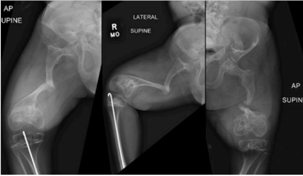 . Images of an 8-year-old patient with severe type III OI who has experienced multiple femur fractures over time that were treated without surgical fxation. Note the signifcant deformity, nonunion, and developing pseudoarthrosis. OI, osteogenesis imperfecta.
Figure 2. A 12-year-old male with type I OI presenting with a classic olecranon
avulsion fracture. The injury happened when he extended his arm to break his fall. Notably, the patient's father also has type I OI and experienced bilateral olecranon avulsion fractures approximately one year apart when he was around
the same age as his son. PLEASE REFERENCE LEARNING HOW TO FALL
Figure 3. 12-year-old girl with OI with K-wire fixation and a cerclage construct
using a telescopic intramedullary rod. OI, osteogenesis imperfecta.
OI, osteogenesis imperfecta
OI in children with moderate to severe disease, and special consideration is needed in these cases.Certain special considerations in OI bone should not be overlooked. First, to maximize the bone's ability to heal, a saw should not be used to prevent heat necrosis. Depending on the bone's brittleness, osteotomies can be performed with a rongeur alone or using a multiple drill hole technique, then completed with a rongeur or osteotome. To preserve the soft tissue envelope and healing periosteal sleeve, a percutaneous osteotomy should be performed whenever possible, except in the distal
humerus, because the radial nerve is inevitably draped over the apex of the varus deformity.
Traditionally, the goal of intramedullary nail systems for OI patients is to provide protection of the entire bone length and decrease stress risers at the ends of hardware. If needed for length or rotational stability or nonunion treatment, a small locking plate with unicortical screws may be considered as a supplemental construct in conjunction with an intramedullary device [26]. Supplemental plates are not the answer to all OI surgical difficulties, and they should be used judiciously and not routinely. Despite using the plates only in conjunction with intramedullary devices, there can still be complications such as fracture below the plates and loosening of screws. One study noted that the advantages are primary rotational stability and earlier weight-bearing in these challenging cases [29]. Another study, which utilized supplemental plating due to inadequate intramedullary (IM) fixation, rotation stability, or persistent cortical gaps, found many complications including screw pullout and peri-implant fractures, both before and after plate removal.
One other important concept is to avoid accepting a starting point for any intramedullary device that is not aligned with the long axis of the bone as the starting point for the first rod will be the starting point for the life of the bone. In the femur, if the starting point is not just inside the greater trochanter, you predispose the proximal femur to drift into varus over time. Another important aspect is to not insert a rod that is too large into the bone. If you do, the rod will take on too much of the force, resulting in stress shielding of the bone and significant bone loss, which is hard to recover from.
Telescopic and non-telescopic intramedullary rodding Telescoping rods are widely regarded as the preferred fixation method for growing children. A multicenter study demonstrated longer implant survival with telescoping rods (5.8 years) than with static rods (4.0 years), as well as fewer reoperations during long-term follow-up [35]. Nontelescoping rods can be utilized in patients with OI and are
most often utilized in bones that are too small to accommodate a telescoping rod, or in patients who are skeletally mature. A case series of nontelescoping threaded (SLIM nail, Orthopaediatrics, Warsaw, IN)
versus nonthreaded rods (examples are rush rods and k-wires) found that the nonthreaded rods demonstrated six rod prominences requiring revision and three rod migrations, while the threaded group had no prominences or migrations. The relative risk of complications in the nonthreaded group was 4.67 (95% CI: 1.47-14.82) compared to the threaded group [29]. A prospective multicenter study of seventy-eight children treated with telescoping nails reported fracture union in 97.4% of cases and deformity correction in 91% at one year. Functional outcomes improved significantly, with a complication rate of 23%
Although telescoping rods are now the mainstay of treatment in both fracture and deformity correction, one 2024 study examined sliding double flexible intramedullary nails (SDFIN) for fracture treatment and deformity correction. They placed two straight flexible titanium nails per bone, one antegrade and one retrograde. They had eleven patients with thirty-three femurs and tibias treated. They found similar rates of union and revision rates compared to published studies on FD rods.
The conclusion was that SDFIN is a viable and cost-effective alternative treatment, especially in resource-limited settings [37]. Several authors from Europe reported on a new telescopic nail sys-
tem introduced in 2022, the Rodeo by Orthofix® (Lewisville, TX). One surgeon in Germany presented his results in 2025 in 5 patients, twelve nails (9 femurs, 3 tibias). They found typical OI bone healing with no loss of correction, and only one femoral endcap loosening that required reoperation. They reported that there are some technical advantages of this new nail system over prior systems they had previously used and noted that they preferred to do the femurs retrograde rather than
anterograde.
Humerus
The humerus can be treated with an intramedullary telescoping nail if the bone is large enough, with either antegrade (Fig. 5) or retrograde (Fig. 6) approaches
FIG 5 FIG 6
There is a significantly increased risk of nonunion in the distal humerus and there are many case reports in the literature on treatment. A recent publication described the treatment of nonunions with intramedullary nails and supplemental plating with possible adjunct bone grafting. There is significant variation in the success of this treatment. In settings of frank pseudoarthrosis of the distal humerus, one should consider nonoperative management if the epiphysis in a center–center position. OI, osteogenesis imperfecta.
If the patient has hand function [38]. Care also should be taken to protect the radial nerve as it is almost always at the site of nonunion or the distal humerus varus deformity Oder et al. [39] published a retrospective study on upper-extremity deformities and functional impairments in 14 patients with OI, aged 8-73 years. They evaluated X-rays, clinical function, and range of motion (ROM). Radiographic findings revealed diverse deformities, with more severe deformities and radial head dislocation correlating with compromised ROM and worse function. They also found greater joint kinematic deviations and slower task execution times. They concluded that functional impairment in daily activities among OI patients is variable and largely depends on the severity of upper-extremity deformity, emphasizing the need for larger multicenter studies.
Femur
Treatment of femur fractures or femoral deformities in children with OI requires straight nails, which must be placed just inside the greater trochanter. One study reported on 150 femurs with an
average follow-up of 8 years and found that the entry site was medial to the tip of the greater trochanter in 26% of patients, at the tip of the greater trochanter in 43%, and lateral to the greater
trochanter in 31%. They had one patient who developed avascular necrosis (AVN), but this occurred after the patient sustained a displaced FNF, indicating that placing the rods just inside the greater
trochanter is not a risk factor for AVN [29]. When the rod is not placed just inside the greater trochanter, there is a risk as the child grows of the rod may migrate laterally, which can result in recurrent proximal femoral varus deformity, possible recurrent fractures, or nonunion
.
FIG 8
A challenging aspect of placing the telescoping rod in the femur is deciding how deep to insert the female nail into the greater trochanter. A series of 136 femoral roddings showed that when the rod is threaded only into the apophysis, the risk of distal migration is 4%, compared to 12% when the threads extend across the apophysis. This is important because rod migration into the
Type III OI patient with distal humeral shaft fracture and varus deformity treated with an antegrade FD nail. FD, Fassier–Duval; OI, osteogenesis imperfecta..
Figure 6. a) Example of retrograde male nail-only fixation of the humerus. b) Preoperative humeral deformity. c) Postoperative deformity correction with a retrograde FD nail. FD, subtrochanteric region increases the risk of subtrochanteric femur fractures and complicates future rod revision surgeries.
Figs 9
Tibia
One recent study presented on 102 OI patients who had undergone 428 tibial telescoping rod
surgeries. They evaluated the location of the male component of the FD rod in the distal epiphysis. They found the overall failure rate for telescoping was 46%, and that rods placed anteriorly or posteriorly had higher failure rates (58% and 56%, respectively) than centrally positioned rods (35%; P < .001). Similarly, rods placed in the lateral third failed to telescope in 70% of cases compared to 46% in the medial third and 35% in the central position (P < .001), Fig. 11. They concluded that center–center alignment of FD rods in the distal tibial epiphysis is ideal to maximize the longevity of the
rod and its ability to telescope.
FIG 10
Nonunions
Nonunion, whether due to delayed bone healing, atrophic, hypertrophic, or infected, adds a significant layer of complexity to bone surgery in OI. Several clinical factors such as vitamin D levels, nutritional
status, bisphosphonate use, or other medications, can influence a patient's healing potential. Nonunion is often associated with repeated fractures at a progressively deforming site [40]. The most common sites are the proximal femur, mid-shaft tibia, and distal humerus as shown in Fig. 12.
FIG 12
One study employed the RUST (Radiographic Union Score for Tibial Fractures) scoring system to objectively evaluate fracture and osteotomy union in OI patients [41].The RUST has demonstrated excellent inter-and intra-observer reliability when assessing bone union with an intra-medullary device in place. Even with radiographic criteria, some patients are symptomatic and require surgical intervention, while others a locking plate is directly under the nerve during this nonunion surgery.
Left femur osteotomy with FD nail placement through the tip of the greater trochanter at age 5. b) Two years postoperation, showing slight lateral migration of the rod. c) Four years postoperation, showing continued lateral migration of the rod and subtle development of proximal varus and incomplete bony
union at the previous osteotomy site.
Some patients show no clinical symptoms despite similar radiographic findings.Agarwal et al. [40] reviewed 44 patients and identified 9 nonunions (18%)-4 atrophic, 2 with a gap, and 3 hypertrophic. Interestingly, they found that radiographic nonunion at osteotomy sites was often asymptomatic, whereas postfracture nonunion generally needed surgery. The location of nonunion and OI type influence the severity of symptoms related to the nonunion.
Timing of pamidronate infusions is crucial for perioperative fracture and osteotomy care. Munns et al. [41] reviewed 197 fractures and 200 osteotomies, finding more delayed healing after osteotomies when
pamidronate was started before surgery. Furthermore, they observed that during pamidronate treatment, older age and tibial osteotomies were independent risk factors for delayed healing. Concerning fracture
healing, after accounting for age, they found no significant differences with or without pamidronate; instead, better mobility status was the strongest independent predictor of delayed healing. Recent advances in surgical technique and perioperative management have reduced rates of delayed healing. Anam et al. incorporated strategies such as avoiding the use of a power saw, withholding bisphosphonates for 4 months postosteotomy, and transitioning to zoledronate—all of which
decreased the nonunion rate in 139 femurs and 112 tibias from 72% to 42% [42]. Additional approaches could include autograft, allograft, synthetic substitutes, or bone morphogenic protein
Lastly, a study out of Hong Kong looking examining time to union of fractures found that children with WNT1 mutations (type XV OI) had a slower time to union (57 ± 9 days) compared to children with COL1A1 variants (51 ± 9 days), both of which were slower than those in a normal control group (35 ± 9 days, P < .001) [23].
Spine
Spinal deformity is common in OI and may affect up to 80-90% of patients with severe disease [42–44]. Kyphoscoliosis is the most Figure 9. a) Patient with type IV OI who had osteotomy and FD rodding at age 4; note that no threads cross the greater trochanteric apophysis at the time of the
initial procedure. b) As the patient grew, the rod remained in the same position, and once the rod was telescoped about 50% of the femur's length, a straightforward exchange nailing was performed. The patient was able to have the procedure done as an outpatient with no weight-bearing restrictions postoperation. c) One month after the exchange. d) Patient with type IV OI who had threads completely crossing the apophysis at the initial procedure at 4.5 years old, now at 8 years old. e) With the rod having grown down, it migrated laterally, with mild proximal varus deformity. FD, Fassier–Duval; OI, osteogenesis imperfecta.
Figure 10. A 6-year-old female with type IV OI who developed proximal femoral overgrowth, causing the female portion of the rod to be located in the subtrochanteric region, which resulted in a subtrochanteric femur fracture. OI, osteogenesis imperfecta. frequent manifestation, although spondylolisthesis and craniocervical abnormalities such as basilar invagination (BI) can also occur.
Curve progression correlates with disease severity and bone density [45]. Children with rare genetic variants such as IFITM5 or WNT1 often demonstrate more severe scoliosis compared with those with COL1A1 and COL1A2 variants. Leg length discrepancy, BMD, and age at first drug use (all proxies for increased OI severity) were correlated with increased severity of scoliosis outcomes. In a study by Chen et al. [46], out of 290 patients, 59 had severe scoliosis (>50-degree curves), and 25 of these
patients had undergone posterior spinal fusion. These patients may be able to attempt bracing, but consideration should include both ventilatory restriction and the understanding that a brace is meant for support and comfort, rather than to prevent curve progression. Ultimately, some of these patients may progress to the point of requiring surgical intervention, often performed for curves >40-50◦ . When cor-
recting the scoliosis, implant density can help distribute bony stress, and Figure 11. a) Patient with severe recessive OI. Right tibia with male nail in the center position on the AP view at the time of surgery. b) Left tibia of the same patient with the male nail in the lateral epiphysis. c) Eighteen months postoperative, although both rods are no longer telescoping, the right rod telescoped longer than the
left, and the right has maintained better deformity correction, highlighting the importance of the center–center position of the male nails in the distal tibial epiphysis during the initial procedure. OI, osteogenesis imperfecta.
Figure 12. An 8-year-old male with symptomatic mid-shaft tibia nonunion treated with revision FD nail and locking plate. He proceeded to union and was asymptomatic at 3 months postoperative.the screws can be augmented with cement to improve pull-out strength [43,42]. The correction itself should be much gentler, with a goal to achieve deformity stabilization to prevent progression rather than to
maximize correction of the Cobb angle [44]. Pelvic instrumentation can be considered, particularly for the nonambulatory patients with high pelvic obliquity.
An abstract presented at ICCBH in 2024 evaluated 148 children with OI including 101 with scoliosis and 47 without. The study found that children with scoliosis had lower self-image (3.31 vs 3.7, P = .003) and mobility (3.13 vs 3.66, P = .015). Greater curve magnitude was linked to increased pain and anxiety but was inversely related to function, self-image, and total Scoliosis Research Society (SRS) score. Patients who underwent surgery reported higher treatment satisfaction scores (4.41
vs 3.43, P = .002) compared to patients with scoliosis and a Cobb angle >50◦ who did not have surgical treatment.
A single-center prospective study of 35 patients who underwent posterior spinal fusion was presented at the 15th International Conference on OI (Fig. 13). The cohort had a mean follow-up of 5.5 years with a
mean age of surgery of 12.2 years. They found a 56% correction rate for curves, with improvements in SRS-22 total score (3.6-4.1, P = .002), mobility, and patient-reported fatigue. They reported two complications: one patient with proximal junctional kyphosis and another with
an intraoperative femur fracture.
Scoliosis can appear in some children with OI at an early age. A recent multicenter database study evaluated growth-friendly spinal instrumentation in fifteen patients with OI, with a mean age of 7.3 years, and found a 35% correction of the coronal curve after the initial surgery. Children with OI had a significantly higher risk of proximal anchor failure (53%) compared to 20% of children with idiopathic curves undergoing growth-friendly spinal fixation. OI children who underwent preoperative halo-traction experienced greater length gains and more correction of the coronal curve at final follow-up.
Basilar invagination occurs in up to 40% of individuals with OI and may cause neurological symptoms from brainstem compression [43,47]. A detailed history and clinical examination can reveal signs and/or symptoms of BI. A lateral cervical radiograph is recommended for all OI patients [47], and it can be diagnostic for invagination, although details of the craniocervical junction may be difficult to visualize in some patients. Further imaging may include a magnetic resonance imaging (MRI) as shown in (Figs 14 and 15), to assess spinal cord changes, along with a CT angiography for surgical planning and anatomical mapping. Patients with symptomatic BI should undergo decompression, distraction with halo traction, and fusion to the occiput.
Lastly, spondylolisthesis occurs more frequently in these patients than in the general population [47,49–51]. It can be detected through standard scoliosis screening imaging. A study published in 2011 found a spondylolysis prevalence of 8.2%, with 7 out of 9 patients having type III OI, and a spondylolisthesis prevalence of 10.9%, involving 4 out of 12 with type I, 4 out of 12 with type III, 2 out of 12 with type IV, and 2 with unknown types. The same group was evaluated ten years later, revealing 15% with spondylolysis and 18% with spondylolisthesis, none requiring Figure 13. 12-year-old female with type III OI and preoperative 100-degree thoracolumbar curve. She underwent posterior spinal fusion, with cement augmentation noted at the proximal and distal screw levels. OI, osteogenesis imperfecta.
FIG 14
<---
FIG 15
--->
. A 15-year-old female with type III OI. Note the dens within the skull base and compressing the brainstem; hydrocephalus is also present. OI, osteogenesis imperfecta. Figure 15. same patient 8 years postoperative from decompression and craniocervical fusion. surgical fixation or repair.
Additionally, 68% of these patients also had scoliosis [32]. For patients with concurrent kyphoscoliosis, it is advisable to include the spondylolisthesis in the construct, especially in
wheelchair-bound patients with pelvic obliquity. In cases of isolated spondylolisthesis without scoliosis, decompression and stabilization may be necessary if symptoms develop, although this is rare in the authors’ experience. One important factor in considering stabilization in
these children is bone quality as poor bone quality can lead to peri-implant stress injury [52]. Therefore, preoperative optimization of bone density is recommended.
Conclusion
Caring for children with OI is challenging yet rewarding and often involves more than just managing their orthopaedic care. It is important to have a proactive, multidisciplinary team dedicated to the complex care of these children and their families. The Osteogenesis Imperfecta Foundation offers many patient, family, and medical resources on its website. We highly recommend the School-Aged Tool Kit as a resource for both families and physicians [53,54]. Ethics approval and consent No identifying patient information as utilized in this review article.